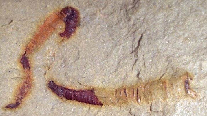
A 550 million-year-old fossilized digestive tract found in the Nevada desert could be a key find in understanding the early history of creatures on Earth.
Over a half-billion years prior, life on Earth was involved in simple ocean organisms unlike anything living in the present oceans. At that point, starting around 540 million years prior, creature structures changed dramatically.
During this time, precursors of numerous creature groups everyone realize today showed up, for example, crustaceans and worms, yet for years researchers didn’t have the foggiest idea how these two unrelated communities of creatures were associated, as of recently. An investigation of tubular fossils by researchers led by Jim Schiffbauer at the University of Missouri gives proof of a 550 million-year-old digestive tract—one of the oldest known examples of fossilized interior anatomical structures—and uncovers what researchers believe is a potential response to the question of how these creatures are associated. The investigation was published in Nature Communications.
“Not only are these structures the oldest guts yet discovered, but they also help to resolve the long-debated evolutionary positioning of this important fossil group,” said Schiffbauer, an associate professor of geological sciences in the MU College of Arts and Science and director of the X-ray Microanalysis Core facility. “These fossils fit within a very recognizable group of organisms—the cloudiness—that scientists use to identify the last 10 to 15 million years of the Ediacaran Period, or the period just before the Cambrian Explosion. We can now say that their anatomical structure appears much more worm-like than coral-like.”
The Cambrian Explosion is broadly considered by researchers to be the point in the history of life on Earth when the ancestors of numerous creature groups everyone realize today developed.
In the study, the researchers utilized MU’s X-ray Microanalysis Core facility to adopt a one of a kind investigative strategy for geological science—micro-CT imaging—that made a digital 3-D picture of the fossil. This strategy enabled the researchers to see what was inside the fossil structure.
“With CT imaging, we can quickly assess key internal features and then analyze the entire fossil without potentially damaging it,” said co-author Tara Selly, a research assistant professor in the Department of Geological Sciences and assistant director of the X-ray Microanalysis Core facility.
Disclaimer: The views, suggestions, and opinions expressed here are the sole responsibility of the experts. No Opinion Bulletin journalist was involved in the writing and production of this article.